

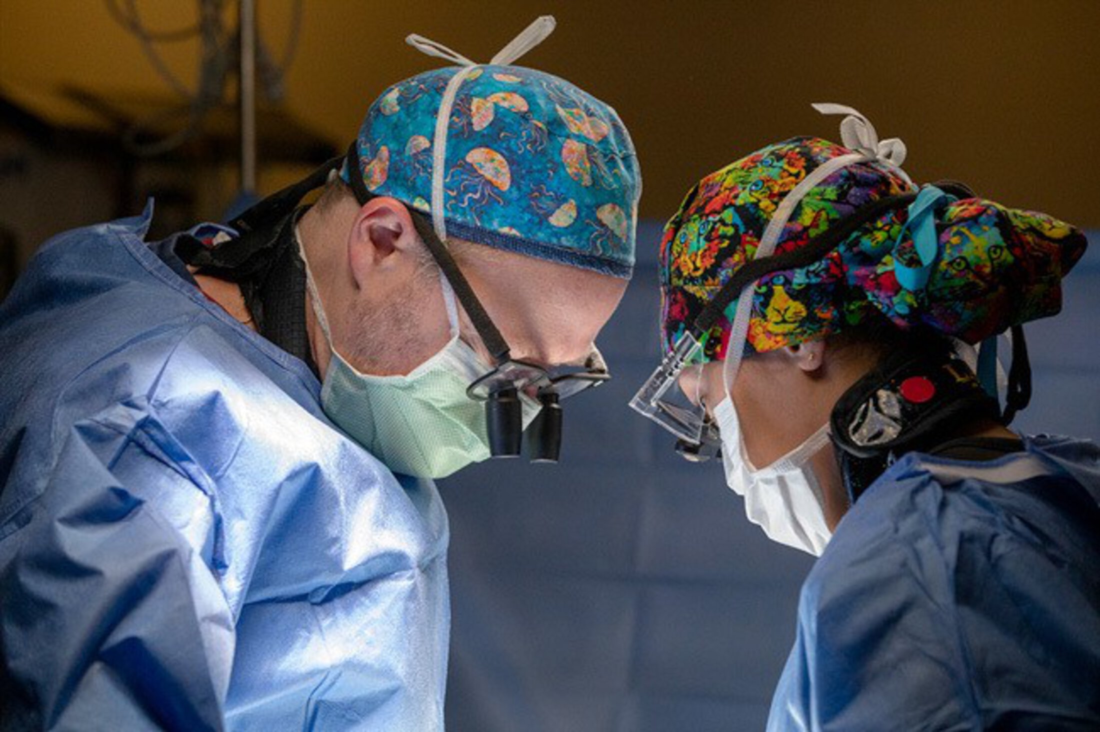
Surgery to remove a herniated thoracic disc is among the most technically demanding procedures in spine surgery, requiring surgeons to operate millimeters from the spinal cord in an area where even minor missteps can have devastating consequences.
For decades, spine surgeons accepted those risks as part of the procedure. But in 2016, Stephen Kalhorn, M.D., a professor of neurosurgery at the Medical University of South Carolina, began to wonder whether there might be a better way.
“I spent all day trying to get the herniated disc out through the chest, and I had to stop because I was concerned. I could cause a hole in the spinal canal and cause a spinal leak, or worse,” Kalhorn said. “After closing and reassessing the approach, I went home that night thinking, ‘How am I going to get the rest of that out of there?’”
After closing and reassessing the approach, I went home that night thinking, ‘How am I going to get the rest of that out of there?’
The next day, Kalhorn and his team approached the disc, which was also calcified, from the back (posterior) and slid it out easily, leading Kalhorn to ask himself: “Why the heck don’t we do this every time?”
In truth, there were good reasons to avoid approaching the thoracic disc from the back, primarily because its close proximity to the central nervous system posed risks of spinal cord damage and paralysis. For decades, this approach was considered too dangerous to perform, Kalhorn explained.
To support the posterior approach, Kalhorn recently published a study in the Journal of Neurosurgery (JNS): Spine– a medical account of 108 successful cases that he’s performed. The accounts span 10 years of Kalhorn’s practice at MUSC and represent the largest known case series on thoracic spine surgery.
No longer unorthodox
The traditional method of removing herniated discs from the front requires a large incision through the chest called a thoracotomy. Sometimes a rib needs to be removed, and a thoracic surgeon will help to access the thoracic spine.
“The procedure involves a chest tube, ICU stay, and significant pain and risks like a pneumothorax or major bleeding,” said Kalhorn.
Notably, this anterior approach was first pioneered, in part, at MUSC by physician Phanor L. Perot Jr., M.D., explained Kalhorn. Perot was a towering presence in academic neurosurgery and considered “South Carolina’s father of neurosurgery.”
Utilizing this novel posterior approach, Kalhorn accesses the spine from the back, typically creating an incision of only one to two inches, avoiding the need to open the chest.
“It’s now essentially an outpatient procedure. Patients don’t need ICU care, chest tubes or extensive pain management,” he said.
It’s now essentially an outpatient procedure. Patients don’t need ICU care, chest tubes or extensive pain management.
In the 2026 JNS publication, Kalhorn and his co-authors demonstrated that posterior thoracic disc surgery can be performed safely with newer technologies, such as an ultrasonic device called Sonopet, which extracts calcified disc material.
“Thoracic discs are often calcified, which makes them difficult to remove, but this tool allows us to carve them out with minimal vibration near the spinal cord,” said Kalhorn.
Before finishing the surgery, intraoperative ultrasound is used to ensure that the disc is completely removed, and the spinal cord is decompressed.
“That’s why we call it ‘ultrasonic spine surgery,’” noted Kalhorn.
Kalhorn said another advantage of the posterior approach is that it can work at any level or on any vertebra of the thoracic spine.
“The traditional thoracotomy approach can’t access certain upper thoracic areas due to anatomical constraints like the scapula. With this technique, we can remove discs of any size, even large, calcified ones."
Advancing innovation, inspiring the next generation
As an academic surgeon, Kalhorn recognizes the importance of inspiring the next generation of surgeons in novel approaches to spine surgery.
Kalhorn’s embrace of a posterior approach to thoracic disc surgery has inspired his trainees, said co-author Brian Saway, M.D., a sixth-year neurosurgery resident at MUSC and future spine surgeon.
“Dr. Kalhorn developed a way to treat this pathology without completely deconstructing the spine or placing screws and rods,” he said.
Saway said these less-invasive tactics can help to minimize risk.
“Even the smallest manipulation can injure the spinal cord,” he explained. “The technique uses ultrasound for real-time intraoperative imaging and an ultrasonic aspirator to carefully shave away bone while preserving spinal stability.”
For Saway, the technology reflects a broader evolution in spine surgery – one that improves outcomes and reduces risk.
“The ideal approach may combine the principles of traditional surgery with newer minimally invasive techniques to create a harmonious balance – the ‘perfect surgery,’” said Saway.
Together, these innovations are expanding treatment possibilities for patients who have historically faced limited or especially high-risk surgical options.
“These are highly complex cases, and patients benefit from evaluation at academic health centers like MUSC with experience in advanced spine surgery techniques,” Kalhorn said.
“For some patients, these types of specialized approaches can make an important difference in both safety and outcomes, which is why they’re often referred to us.”
For some patients, these types of specialized approaches can make an important difference in both safety and outcomes, which is why they’re often referred to us.
Stephen Kalhorn, M.D., FAANS
- Brain & Spine Cancer
- Neurosurgery
- Spine Surgery
- Mount Pleasant, SC