
Catching Alzheimer’s disease early – maybe even before it causes any problems – could allow patients and their loved ones to learn what may lie ahead and take advantage of treatments while there’s still time. The same might be true for another problem plaguing some people as they age: cardiomyopathy, a disease that can enlarge and/or thicken the heart and lead to heart failure.
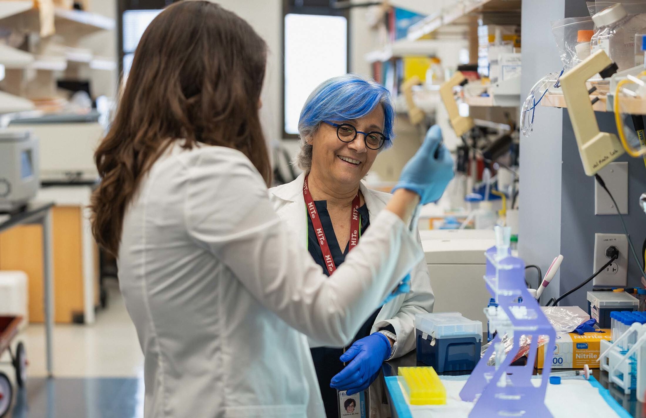
If the idea of pairing the brain and heart possibilities surprises you, you probably haven’t heard about the groundbreaking work of the del Monte Lab at the Medical University of South Carolina. Its leader, cardiologist Federica del Monte, M.D., Ph.D., discovered years ago that there are plaques and tangles in the hearts of people with cardiomyopathy and heart failure just as there are in people with Alzheimer’s disease. “Exact same thing,” she said.
Her team’s work has built on that finding over the years, seeing cardiomyopathy and heart failure as “the Alzheimer’s disease of the heart.” Now, del Monte is leading a study aimed at finding new biological markers, or biomarkers, and other ways to diagnose and monitor brain and heart disease earlier than is currently possible.
The study
Her study will focus on three groups:
-People who have heart failure with preserved ejection fraction.
-People with Alzheimer’s disease.
-People with neither condition who will serve as the control group.
The researchers will collect blood and urine samples to look for biomarkers. The study participants will also get:
-Physical exams.
-Cognitive tests.
-Electrocardiograms, or EKGs, to record the heart’s electrical activity.
-Echocardiograms, also known as echos, which use sound waves to create moving images of the heart.
Del Monte said the work was made possible through a generous anonymous donation. “It allows us to purchase very specialized equipment that will allow us, for the first time, to compare whether diagnosing amyloid in the heart and hopefully in the future in the brain with an echocardiography is as specific and sensitive as PET imaging.”
PET stands for positron emission tomography. PET imaging “uses small amounts of radioactive materials called radiotracers or radiopharmaceuticals, a special camera and a computer to evaluate organ and tissue structures and functions. By identifying changes at the cellular level, PET may detect the early onset of disease before other imaging tests can,” according to RadiologyInfo.org.
Another question
Another question del Monte’s team is trying to answer is where amyloid formations show up first – in the heart or the brain? “It is possible that it doesn't only start in the brain. Functionally, at least in mouse models, the heart shows signs of accumulation of plaques and tangles earlier than the brain,” she said.
If that holds true in people, it could have a big impact. “We could diagnose Alzheimer's ahead of time with an echocardiography. We could say that there are those plaques and tangles in the heart as in the brain, where we just don't see the cognitive symptoms while we see the cardiac signs. It might be possible to predict Alzheimer's in some patients and intervene early because the intervention to the heart and the brain can be shared. Possibly. We don't know yet. We need to run the study,” del Monte said.
The study involves two initial visits with four-year and eight-year follow-ups.
Separate but related research
Del Monte is also involved in work focused on experimental antibodies designed to treat heart failure. One has been patented and tested in the lab but not in people. “It clears the plaques and improves the molecular signs that are associated with heart failure. Cardiac function is also improved.”
But there are side effects. “So we’re working on different types of antibodies that we can’t describe yet,” del Monte said.
Hybrid heart/brain clinic
If these antibodies hold up to scientific scrutiny, the study could become part of a hybrid heart/brain clinic del Monte envisions for MUSC. “So my long-term goal is to establish a clinic where neurologists and a cardiologist, a physical therapist and a psychologist work together with patients,” she said.
“The patient sits in the room. The doctors rotate; they do their assessments. The patient goes home. Then, the healthcare professionals sit in a room, discuss the case and make a plan.”
You can keep up with the del Monte Lab’s work through its website.